
Dystocia in mares, although relatively uncommon, represents a true veterinary emergency requiring rapid assessment and intervention to ensure the survival of both mare and foal. Prompt recognition and correction of abnormal fetal positioning or obstructed delivery are essential. Delays beyond 20 to 30 minutes from the onset of stage II labor are strongly associated with increased morbidity and mortality rates for both mare and foal. This case report reflects on the management of an equine dystocia attended in a clinical setting, highlighting the importance of coordinated veterinary response and immediate obstetrical correction. Following successful delivery, intensive neonatal nursing care played a pivotal role in the foal’s recovery. Core interventions included thermoregulation, assisted colostrum intake, monitoring for passive transfer of immunity, and regular vital sign surveillance. Veterinary nurses were instrumental in identifying early signs of neonatal maladjustment and coordinating supportive care in collaboration with the attending veterinarian. This case reinforces the necessity of not only timely obstetrical intervention but also structured, evidence-based nursing protocols post dystocia to optimize neonatal outcomes.
Take-Home Points
- Reported incidence of dystocia in mares is less common than in other species and is as low as 4% to 8%; when it occurs, it is a true emergency.
- Early, decisive action is critical: Manual correction should begin within 15 minutes if there is no progress.
- Detailed protocols for neonatal intensive care show that early and continuous intervention by trained staff is vital.
In the early hours of a winter’s morning, an 11-year-old, approximately 600-kg (1323-lb) chestnut mare was presented to the equine hospital for emergency assessment of possible dystocia. The mare had been in labor for several hours, and the foal was abnormally presented with only 1 hoof visible. Dystocia in mares is defined as any abnormal or difficult birth resulting from either maternal or fetal causes. It is relatively uncommon in mares, occurring in approximately 4% to 8% of all foalings.1 When it does occur, it is often caused by fetal malposition and requires immediate intervention (within 15 minutes).2,3
The mare was in a panicked state, constantly getting up and down and showing signs of discomfort and increasing pain observed as tail flagging, pawing, sweating at the flanks, and generalized restless behavior. After administration of 5 mL xylazine (100 mg/kg) IV and 6 mL methadone IV (10 mg/kg), the mare then became recumbent and sedate enough to allow veterinary team members to manipulate and deliver the foal via an assisted vaginal delivery.
Assisted vaginal delivery involves aiding the mare in expelling the fetus per vagina through manual correction of fetal malposition or traction on the fetus without requiring general anesthesia or surgery.4 The mare recovered uneventfully from the sedation and expelled the complete placenta at time of birthing.
Presentation and Physical Examination
The chestnut colt was born with a spontaneous heartbeat and normal respiratory rate. The foal was initially slow to sit in sternal recumbency and showed abnormal behavior, typical of a dummy foal. Dummy foal syndrome, also known as neonatal maladjustment syndrome or hypoxic ischemic encephalopathy, is a common reason for admittance to veterinary hospitals and is a potentially life-threatening condition affecting newborn foals. These foals are typically born at full term but show neurologic abnormalities within the first few hours to days of life.5 Early and continuous intervention by trained staff is vital.6 The foal also showed signs of hyperresponsive and frantic behavior; this was interspersed with periods of somnolence and an inability to stand (FIGURE 1).

FIGURE 1. Chestnut colt in sternal recumbency receiving intravenous supportive therapy and supplemental oxygen therapy.
On initial assessment, the foal’s limbs did not look contracted, mucous membranes were pink and moist but slightly injected, heart rate was 124 beats per minute (bpm), and respiratory rate was 30 breaths/min (TABLE 1).

Initial blood work showed normal hematology with a mild increase in creatine kinase (CK) of 2000 IU/L. Normal neonatal CK is approximately ≤ 1000 IU/L. CK concentrations are often markedly elevated in foals born after dystocia due to soft tissue trauma and ischemia.8
An entropion of the left lower eyelid was noted. An entropion is an inward rolling of the eyelid margin, most commonly the lower lid, resulting in contact of eyelashes or periocular hair with the cornea. Acquired or secondary causes of entropion include dehydration or emaciation.9
Treatment
Supportive Therapy
A 1-L Hartmann (lactated Ringer’s solution) bolus was administered intravenously; instant improvement of the entropion was observed.
1 mL of diazepam (5 mg/mL) was administered intravenously to facilitate the safe and aseptic placement of an Arrow 16-gauge over-the-wire IV catheter (Teleflex) in the left jugular. The umbilicus was clamped with a plastic umbilical clamp, and supportive intranasal oxygen therapy was started at 5 L/min while a stall was prepared.
Supportive fluids consisted of a Hartmann bolus of 100 mL/hr IV and 10% glucose at 150 mL/hr IV to help support metabolic function and provide necessary energy for cellular function and overall health.
Feeding
Due to a poor suckle reflex, a nasogastric feeding tube (NGT) was placed. Correct tube placement was confirmed by physical external palpation of the esophagus and listening for air movement (gurgling). A ring of elastic adhesive bandage was gently placed around the foal’s muzzle, and the NGT was then secured to the ring to ensure the tube stayed securely in place and minimized the risk of displacement.
The mare’s colostrum was collected and was of good quality, having a Brix reading of 28%. Good-quality colostrum should have an immunoglobulin G (IgG) content of ≥ 30 g/L; this equates to a specific gravity of ≥ 23% on the colostrum refractometer scale.10
When milking the mare, monitoring of milk production and output is recommended to ensure her nutritional requirements are met.
Two hourly treatments required the veterinary nurse to perform milk collection from the mare and then feed this to the foal via the indwelling NGT. The foal continued to receive 150 mL q2h of colostrum for the first 12 hours. At 8-hour intervals, feeds were increased by 50 mL.
Positioning for Recovery
Because the foal was unable to stand unaided, it was placed in a foal sternal pad. Positioning the foal in sternal recumbency is the ideal resting or recovery position for most down foals as it optimizes ventilation/perfusion of the lungs, facilitates swallowing and nursing behavior, can minimize the risk of aspiration and hypostatic pneumonia, and minimizes the risk of pressure sores and muscle damage.11
Repositioning the foal may require folding techniques or the help of a second person. This requires the staff to stand the foal for a very short period, allowing it to barely hold its own weight, and then place it back down in sternal recumbency, rotating the hind legs from left to right every 2 hours.
During the repositioning phase, it is extremely important that the foal be carefully examined for any early signs of heat in the joints as this could be indicative of possible bed sores or joint infections. Every time the foal is rotated, the bedding should be cleaned and a clean pee pad placed to collect urine. The foal should not be left to sit in urine; this will make the foal cold and can result in bacteria tracking into the umbilicus. Bacterial infection is the leading cause of death in foals in the first month of life.12 In this case, the antibiotic ceftiofur sodium was chosen because it is a third-generation cephalosporin that shows excellent in vitro activity for both gram-positive and gram-negative bacteria.13 Ceftiofur sodium was administered at 5 mg/kg IV q12h for 5 days.
Recovery
Over the next 2 days, the foal continued to receive the mare’s milk via the NGT every 2 hours and was allowed to stand for small periods while being repositioned in the sternal mat. On day 3, the foal started to show signs of improvement, such as kicking its legs and even getting itself out of the sternal support and standing unaided. These are all good signs the foal is becoming stronger and wanting to be more independent (FIGURE 2).

FIGURE 2. Chestnut colt with intravenous catheter and nasogastric feeding tube in place but off continuous supportive fluids.
Feeds via the NGT were reduced by 50 mL every 2 hours as the foal was introduced back with the mare at short intervals, allowing the foal to nurse naturally. On the first introduction, the mare’s udder was completely stripped of milk to limit how much was available to the foal. Consequently, the next 4 times the foal was allowed in the stall with the mare, her udder was only half stripped to make up for the drop in milk fed through the NGT.
By the fourth day, the NGT was removed, allowing the foal to receive all of its nutritional requirements from the mare. IV fluid therapy was abruptly stopped, and the foal was reunited full time with the mare. Observations of nursing, urinating, and the ability to stand and lay down unaided were completed every 2 hours.
The foal continued to improve, with all vital signs being within normal limits (WNL) during regular temperature, pulse, and respiration checks (TABLE 1). The suckle reflex was getting stronger, and the foal attempted to cross nurse. During regular observations on day 4, the foal passed dark-colored, concentrated urine. This is an important clinical sign, especially in the context of dystocia, dehydration, or renal function assessment. Normal urine specific gravity (USG) in neonates is 1.001 to 1.020. The colt’s USG measured 1.028, indicative of dehydration. A half-liter Hartmann bolus was administered following the attending veterinarian’s advice.
After 5 days in the intensive care unit, blood work showed continued infection with a low white blood cell count of 4 × 10⁹ (reference range, 5.3 to 12.2 × 10⁹). There were no immature band neutrophils, and fibrinogen was 3 g/L (reference range, 1.4 to 6.6 g/L). Blood culture results were negative.14
Thoracic and abdominal ultrasonography revealed no significant abnormalities. Due to blood work revealing neutropenia, the left jugular vein containing the IV catheter was also examined; no sign of infection or thrombosis was noted.
Due to funding limitations, the IV catheter was removed and the mare and foal were discharged into the care of the referring veterinarian (FIGURE 3).

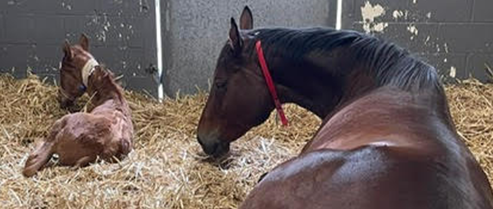
FIGURE 3. Chestnut colt enjoying time in the stall with the mare, after having the nasogastric feeding tube removed.
Outcome
On discharge, the foal’s vital signs were WNL, HR 84 bpm, RR 24 breaths/min, mucous membranes were pink and moist, and temperature was 38 °C (100.4 °F). All extremities were warm, active gut sounds were noted in all 4 quadrants with active borborygmi, there were clear lung sounds, and no abdominal distention was observed. A collected urine sample had a USG of 1.010. The umbilicus was slightly thickened but dry. The young colt was active and nursing well from the mare.
Discharge instructions included monitoring the foal’s temperature every 12 hours for the first 3 days to ensure no fever occurred after stopping the antibiotics, and a new blood sample was to be taken on day 3 of discharge to check white blood cell count differential and fibrinogen.
Follow-up with the referring veterinarian was positive, and the mare and foal did not require any further medical interventions.
Into Practice
Foals born after dystocia are at high risk of hypothermia due to weakness, poor perfusion, and wet coats at birth.
What to do:
- Dry the foal thoroughly and provide external heat sources (e.g., heat lamps, blankets).
- Monitor rectal temperature every 1 to 2 hours.
- Maintain ambient temperature between 25 °C and 30 °C (77 °F and 86 °F) if indoors.
2. Ensure Adequate Passive Transfer of Immunity
Foals born through dystocia may have delayed nursing or poor colostrum intake, increasing risk of sepsis.
What to do:
- Assist the foal in nursing within 2 hours post birth or administer colostrum orally or via plasma if nursing fails. Plasma can be administered intravenously through aseptic placement of an IV catheter or orally if within the first 12 to 18 hours post birth.
- Collect a blood sample between 12 to 24 hours of age to check IgG levels. Blood test (serum IgG) levels below 400 mg/dL often indicate partial failure transfer of passive immunity, and those below 200 mg/dL suggest complete failure transfer of passive immunity.9 If using a rapid, semiquantitative enzyme immunoassay (IDEXX SNAP Foal IgG Test), the same reference ranges can be used.
- Coordinate with the veterinarian to administer IV plasma if failure transfer of passive immunity is confirmed.
3. Implement Frequent, Focused Monitoring
Dystocia increases the risk of hypoxia, trauma, and metabolic disorders. Early signs of deterioration can be subtle.
What to do:
- Monitor and record vitals every 2 to 4 hours:
- Heart rate
- Respiratory rate
- Mucous membrane/capillary refill
- Urination and meconium passage
- Watch for signs of sepsis, limb contracture, or dummy foal syndrome.
- Communicate changes immediately to the attending veterinarian.